Showing posts with label anatomy. Show all posts

Tuesday, July 22, 2014

Increadible - Program for anatomy students

This is payed program, but I post it here to show you that such program already exists.
http://www.teamlabbody.com/3dnote-en/story/index.html

Its having quite high quality images and 3D visualization of human body. It can even display changes of muscles, nerves and bones positions during movements of joins.

http://www.teamlabbody.com/3dnote-en/dictionary/blood_vessels/Profunda_brachii_artery/

Here you can see Radial nerve from back


and from front


I hoped somebody could develop such program and it is already here.

Here is some presentation on Youtube:

Tuesday, May 21, 2013

Latest diagram of location of Radial nerve on Humerus

I did new diagram based on study of Radial nerve and humerus bone. This diagram is great because it contains descriptions. This is posterior view (back).

How to read diagram:

You need some image or arm with muscles like those medical images bellow. I looked for some way how to measure distances. I have found points which are approximately same distance from each other. These points are marked by long horizontal arrows filled with darker color. These are borders of the quarters. Use it as a rule, as if you would put 0 on the blue line. Then read it from bottom upwards. This is the way how I measured the distances. Notes in regular font are description if you go upwards. Notes in italic font are made for the case you read the diagram downwards, as in usual direction.

This diagram should be precise. It revealed some little mistakes in my previous estimations. I would dare to discuss this diagram with some medical educated person, but still be aware I am not a medic.


According other schemes of radial nerve, the path can vary. I have seen path going a bit more to right: in the triangular interval it turns right and does not enter tendon of the triceps. In that case, people with radial nerve like this, will be voluntary in the area between Deltoid and Triceps as I simplify to call it "valley". Also I have seen schemas, where tendon is shorter than this one and different schemas having either thinner or wider humerus.

Notes:
brachii - of arm
posterior - back
anterior - front
lateral - side

Note that at 2/4 and 3/4 radial nerve is covered by triceps medial head (great muscle). At the bottom of humerus it is covered by radiobrachialis muscle, which covers just little part of humerus on the left side of lateral view. You can use this diagram commonly with biodigitalhuman.com.

If you use Takate kote and you look for radial nerve path in takate kote, you must imagine that you rotate the humerus a bit towards right. I guess this rotation is about 20-30°, and can affect the look of the image just a bit.

PS:
I add yet two images, there I tried to draw radial nerve according this diagram. I had to change proportion a bit because the humerus diagram was shorter, that model's upper arm.


You can also use this image to see the relation of position of Deltoid-Triceps-Lateral-Head valley and radial nerve located on humerus.

This source contains this image, showing cross-cuts of humerus, which is to show more exactly where the radial nerve (N. radialis) can lie:


 You can see both views at same time: posterior and lateral. Anterior or back view is when you watch from bottom of the images upwards. Lateral or side view from outside to inside you get when you watch from right to left, and the image from profile.

Monday, May 20, 2013

Radial nerve schemes II & references

I found some interesting images.

First from posterior view, displays the radial nerve path in non rotated arm.

From Lippincott Williams & Wilkins Atlas of Anatomy (Tank, Patrick W.; Gest, Thomas R)

Here is anterior view (front view).


The spiral groove is only area where nerve is located on humerus bone. There for fractures leads to damage of the nerve.


To find where the radial nerve begin we need to check muscles, especially teres minor and deltoid:


References: Great sites for anatomy study!

Virtual Medical Centre

Virtual Medical Centre.com - video: Shoulder joint (including humerus articulation/rotation)

Pediatric Orthopedics: Humerus bone - detailed views


Pediatric Orthopedics: Muscle anatomy of Arm


Lippincott Williams & Wilkins Atlas of Anatomy (Tank, Patrick W.; Gest, Thomas R)

Monday, January 7, 2013

Radial nerve schemas I.

Important note: This post is not finished. I have also images of male, where I would like to draw humerus, deltoid and radial nerve.


I have draw this schemas, which could not be 100% exact. This for illustration and it is based on inspection of muscles of the models I tied. I did not have any medical scans to be exact for radial nerve location. This drawings was again good training for my memory and orientation. Do you know what? I had a problem to find how the humerus should look in this position, commonly with ulna bone. I find out, there is one good point to start with. In a normal position, that is the normal position where medical pictures are usual, your elbow is at back, your palm is open to forward of you. As painters are used to make their sketches, some of them start with joint having shape like a cube. This is exactly case of posterior side of elbow (elbow viewed from back). You see 3 points, like 3 vertexes of cube. The last vertex is invisible and is orientated upwards. Just imagine a cube on a back side of elbow. This is a good starting point.

Normally, the posterior side of humerus (and elbow) is located at back. In takate kote alias box tie position, you have it placed to side. That is cca 90° rotation. Remember, what we are viewing, it is posterior view. This is that we often see when we check the position of wraps on riggers photos.

However, when you tie takate kote, and you stay behind back of your bunny, you see the interior side of the arm (arm = brachium, lat.).


My estimation of Radial nerve on Esinem's body

With permission of Esinem I publish this scheme which I made based on the muscles visible on the photo.

Yet I add the original photo of Esinem:


Monday, July 4, 2011

Radial nerve rotation

Note 1: Maybe better word instead rotation should be "pronation", but I am not sure. Pronation means turning back of forearm to front (supposed that hand is down beside body). I have seen a pronation movement on medical software, but this movement seemed to me unlikely, because only forearm turned, but the humerus bone of upper arm did not rotated. So in this article I suppose that humerus bone rotates when you try to give hand in takate kote position.

Note 2: This article concerns only one vulnerable area, that is placed just under Deltoid muscle. Another vulnerable area for TK positions is placed just above elbow, and you can identify it from medical/anatomy pictures.

For those who are interested on radial nerve location (concerning TK safety):

Scheme of Humerus bone - size cca 2 cm:
sizes of the humerus

View on non rotated arm, radial nerve:
"back" view on the arm
"back" view on the arm 2

My schemes to illustrate how much the radial nerve "position changes" during 45° rotation:

Test of rotation of "arm" / humerus bone:


I took a pole and paper and draw the lines on it. According scheme that I have. The red line is radial nerve (a piece of wire that I turned around according scheme).

Rotated and non rotated:

Back side of shoulder would be on left and elbow on right side. These two images I use to paste them in schema, so that the lines that I have drawn, would fix schema.
Area located between black lines no. 2 and no. 3 would seem to me safe for face-down suspension, but because the arm have to be rotated 45° in Takate Kote position, then the radial position moves more up, too close to rope.

Non rotated:


Rotated:


Vertical:

Non rotated - rotated:


The dangers area no. 1


Results of the test:

This demonstration should show how much the radial nerve "changes position". The area from black line no. 2 to no 3. is the area I still speak about.

Result:
If you see the vertical image - from the test is possible to see that the radial nerve appears to be close to right side.

The marked area black lines 2 - 3:
It seems to me that the lower 1/2 is dangerous for face down suspension. In the very edge of the Deltoid it could be really dangerous to have ropes for suspension. Then, in the upper 1/2 of marked area, it could be not so dangerous for F-D susp. but for safety reasons we should to place the ropes above this area.

For side suspension and back suspension the marked area in black lines 2 - 3 would be very dangerous area, where the ropes wouldn't be placed!

What danger?

The danger, that is related with incorrect rope placement or too tight tie, is the possibility that you can damage nerve. Too tight tie can cause nerve compression and this can hurt it. If we speak about radial nerve, which is placed in hand, then the injury causes impossibility to use toe or index finger. It can take few weeks to heal the nerve. To prevent this, we should place rope on body parts, which have muscles strong enough to prevent nerve damage. Rigger has to check ropes correct position and fixation or check it during the bondage play, if the tied person moves with ropes.

Sunday, July 3, 2011

Anatomy/Surgery images for those interested in bondage safety

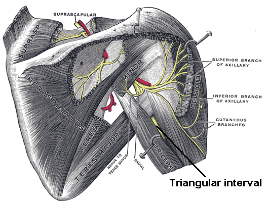
These images are concerning vulnerable areas of limbs. It is good to be concerned in bondage safety. For example in Takate Kote ties we want to know which arteries we could restrain if we would tight the rope in incorrect place or too strong. Especially when some people are doing suspension they should know where the arteries and nerves are because it is possible to damage the nerves or to stop blood circulation in limbs. And because we don"t want anybody get hurt, we study anatomy and all the stuff around bondage safety.

If you click on thumbnails you open images that are hosted on this site - book Surgical Anatomy by Joseph Maclise.

You can find more on internet if you want to look, just search keywords like brachial, axillary, axillae, elbow, wrist. General problem when looking for anatomy images is that we would need special software that is not free, to get 3d images, animations etc, to see were nerves go from and to. Only good surgery images and software for surgery could provide that. Because for good orientation in the space man needs some points like muscles, arteries, veins.

Here is description of inner side of upper arm (not concertning radial nerve)
Upper arm: page

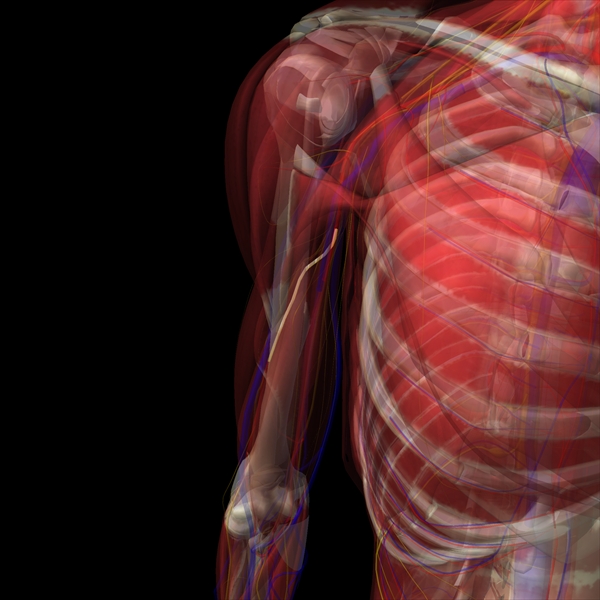
Upper arm & shoulder


Page Plate11
A. Subclavian vein;
   a, the axillary vein;
   a *, the basilic vein, having the internal cutaneous nerve lying on it.
B. Subclavian artery, lying on F, the first rib;
   b, the axillary artery;
  b *, the brachial artery, accompanied by the median nerve and venae comites.
C. Brachial plexus of nerves; c*, the median nerve.
S. Cephalic vein


This is Same page Plate 12 deeoer view to the same front area, when the arm is lifted & supported. You can see the Pectoralis minor (lesser Pectoral muscle: H,K) which is cut. Pectoralis minor lies under Pectoralis major (I), which is removed.

A. Axillary vein, cut and tied;
    a, the basilic vein, cut.
B. Axillary artery;
   b, brachial artery, in the upper part of its course, having h, the median nerve, lying rather to its outer side;
   b*, the artery in the lower part of its course, with the median nerve to its inner side.


Page Plate13

A. Axillary vein
  a - the common trunk of the venae comites, entering the axillary vein.
B. Axillary artery, crossed by one root of the median nerve;
   b - basilic vein,
   a- the axillary vein


Lower arm & upper arm above elbow: 


Page Plate15

H. Radial artery at its middle.
Upper arm inner side:
D. Cephalic vein, with the external cutaneous nerve (left, blue, big arc, outer side)
B. Basilic vein, with the internal cutaneous nerve. (right, blue, inner side)


Same page Plate 16

B. Basilic vein, cut.
C. Brachial artery.
D. Median nerve; d, the ulnar nerve.
F. Origin of radial artery.


Hands


page Plate 17:


B. Median nerve; its branches to the thumb and fin
D. Ulnar nerve; E e e, its continuation branching to the little and ring fingers


Same page Plate 18 
H. Ulnar nerve; h, superficial branches given to the fingers. I


Same page Plate 19
C. End of the radial nerve distributed over the back of the hand, to two of the fingers and the thumb.
D. Dorsal branch of the ulnar nerve supplying the back of the hand and the three ou


Legs - thighs


Page Plate27
f. The middle cutaneous nerve

veins:
d. A common venous trunk, formed by branches from the thigh and abdomen, and joining-- e
e. The saphenous vein.
g. Femoral lymphatic glands.
h. Superficial external iliac vein.
i. Superficial epigastric vein.


page plate28
M. The saphenous vein.N. A tributary vein coming from the fore part of the thigh.


same page Plate 29

I. The femoral vein.
K. The femoral artery.
O. The saphena vein.


page plate 30
N. The femoral artery; n, its profunda branch.
O. The femoral vein
P. The saphena vein


Schemes of veins and arteries
This image is from: http://www.rsdrx.com


Other great sources:
Google translator - for translation of Latin names

http://www.sciencephoto.com/media/114611/enlarge

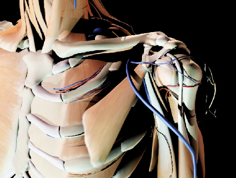
Arteries, nerves and muscles of arm


Radiology site 

http://www.info-radiologie.ch/hip-radiography.php
many of images with descriptions here, but I miss upper arm and side views.
http://www.info-radiologie.ch/anatomy-mri-wrist.php


http://www.imaios.com


You can see there wonderful scans of arms, where also you see the important stuff of muscles (Deltoid, etc)


Also fascinating images (magnetic resonance) of circularity system: saradiology.blogspot.com

And here high quality anatomy schemes: http://imaging.consult.com (sign up free).


Another good site with images of muscles right on bones:

http://www.rad.washington.edu


Really Wonderful site - Magnetic resonance images of inside of human body
http://www.imaios.com/en/e-Anatomy/Limbs