Wednesday, September 4, 2013

Inspiration from Michealangelo Buonarroti

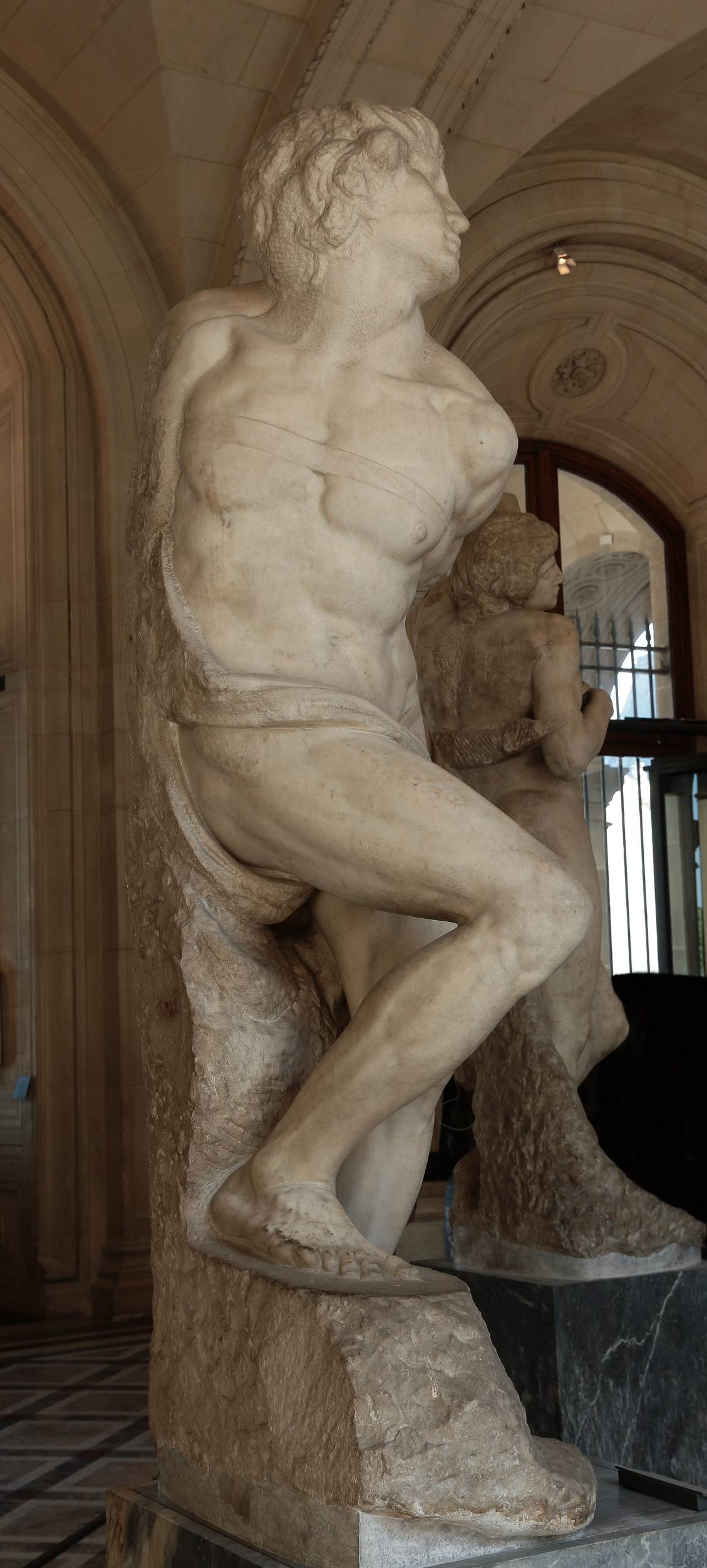
I have found that Michelangelo Buonarroti have created sculpture called Rebellious slave. The sculpture is placed in museum Louvre in Paris. It greatly shows the shapes of muscles of left arm, especially from back view. The arm is placed and rotates in position of Takate kote, hence here is some guide-line for those learning about muscles structure for safety reasons.
http://www.youtube.com/watch?v=h44bnIGogu8


Anyway it was impossible to find any image from back view on net. So if you go to Louvre, please take some image :-) of the Rebellious slave from back.

Tuesday, May 21, 2013

Latest diagram of location of Radial nerve on Humerus

I did new diagram based on study of Radial nerve and humerus bone. This diagram is great because it contains descriptions. This is posterior view (back).

How to read diagram:

You need some image or arm with muscles like those medical images bellow. I looked for some way how to measure distances. I have found points which are approximately same distance from each other. These points are marked by long horizontal arrows filled with darker color. These are borders of the quarters. Use it as a rule, as if you would put 0 on the blue line. Then read it from bottom upwards. This is the way how I measured the distances. Notes in regular font are description if you go upwards. Notes in italic font are made for the case you read the diagram downwards, as in usual direction.

This diagram should be precise. It revealed some little mistakes in my previous estimations. I would dare to discuss this diagram with some medical educated person, but still be aware I am not a medic.


According other schemes of radial nerve, the path can vary. I have seen path going a bit more to right: in the triangular interval it turns right and does not enter tendon of the triceps. In that case, people with radial nerve like this, will be voluntary in the area between Deltoid and Triceps as I simplify to call it "valley". Also I have seen schemas, where tendon is shorter than this one and different schemas having either thinner or wider humerus.

Notes:
brachii - of arm
posterior - back
anterior - front
lateral - side

Note that at 2/4 and 3/4 radial nerve is covered by triceps medial head (great muscle). At the bottom of humerus it is covered by radiobrachialis muscle, which covers just little part of humerus on the left side of lateral view. You can use this diagram commonly with biodigitalhuman.com.

If you use Takate kote and you look for radial nerve path in takate kote, you must imagine that you rotate the humerus a bit towards right. I guess this rotation is about 20-30°, and can affect the look of the image just a bit.

PS:
I add yet two images, there I tried to draw radial nerve according this diagram. I had to change proportion a bit because the humerus diagram was shorter, that model's upper arm.


You can also use this image to see the relation of position of Deltoid-Triceps-Lateral-Head valley and radial nerve located on humerus.

This source contains this image, showing cross-cuts of humerus, which is to show more exactly where the radial nerve (N. radialis) can lie:


 You can see both views at same time: posterior and lateral. Anterior or back view is when you watch from bottom of the images upwards. Lateral or side view from outside to inside you get when you watch from right to left, and the image from profile.

Monday, May 20, 2013

Radial nerve schemes II & references

I found some interesting images.

First from posterior view, displays the radial nerve path in non rotated arm.

From Lippincott Williams & Wilkins Atlas of Anatomy (Tank, Patrick W.; Gest, Thomas R)

Here is anterior view (front view).


The spiral groove is only area where nerve is located on humerus bone. There for fractures leads to damage of the nerve.


To find where the radial nerve begin we need to check muscles, especially teres minor and deltoid:


References: Great sites for anatomy study!

Virtual Medical Centre

Virtual Medical Centre.com - video: Shoulder joint (including humerus articulation/rotation)

Pediatric Orthopedics: Humerus bone - detailed views


Pediatric Orthopedics: Muscle anatomy of Arm


Lippincott Williams & Wilkins Atlas of Anatomy (Tank, Patrick W.; Gest, Thomas R)

Monday, February 4, 2013

Nerve issues (nerve safety)

With conection to bondage we have interests about safety, including possibilities of nerve damage. Therefore we read information about how we can recognize pinched nerve in time.

Article about peripheral neuropathy

This article describes the symptoms of damaged or pinched nerve (they can be similar)
"Patients with peripheral neuropathy may have tingling, numbness, unusual sensations, weakness, or burning pain in the affected area..."
"Neuropathy can present with many differing symptoms, including numbness, pain of different types, weakness, or loss of balance, depending on the type of nerve involved. "

link: peripheral neuropathy

Základní symptomy periferní neuropatie

Vjemové potíže:
necitlivost, neobvyklé vjemové dojmy, brnění, pocit pálení zejména v noci. Dále pak "cizí", studené nohy (tupé, "dřevěné"), slabost

Motorické potíže:
únava, slabost, tíha
křeče (spazmy), fascilukace (pod kůží jsou vidět záškupy svalových vláken, nejedná se však o kontrakci celého svalu)

Odborné pojmy:
Pro zajímavost:
- parestezie je pocit brnění, píchání, svědění či pálení kůže bez trvalých následků
- akroparestezie je parestezie na okrajových částech těla
- hypostezie je snížená citlivost; taktilní hypostezie je snížená hmatová citlivost
- lékaři mluví o rukavicové nebo punčochvité taktilní hypostézii, což je v podstatě akroparestezie
- anestezie (hluboká necitlivost), pallhypestézie (snížení hluboké citlivosti ) a pallanestézie (vymizení hluboké citlivosti)

Závěr
Pokud provádíte bondáž na člověku se zdravými nervy, hlídáte si příznaky neuropatie. Pokud osoba začne mít pocit brnění nebo pálení, ztráty citlivosti nebo tuhnutí končeniny či svalu, je třeba úvaz v dané oblasti uvolnit. Na člověku nemocném polyneuropatií byste měli problém rozlišit, co je příznak polyneuropatie a co příznak skřípnutého nervu.
Monday, January 7, 2013

Radial nerve schemas I.

Important note: This post is not finished. I have also images of male, where I would like to draw humerus, deltoid and radial nerve.


I have draw this schemas, which could not be 100% exact. This for illustration and it is based on inspection of muscles of the models I tied. I did not have any medical scans to be exact for radial nerve location. This drawings was again good training for my memory and orientation. Do you know what? I had a problem to find how the humerus should look in this position, commonly with ulna bone. I find out, there is one good point to start with. In a normal position, that is the normal position where medical pictures are usual, your elbow is at back, your palm is open to forward of you. As painters are used to make their sketches, some of them start with joint having shape like a cube. This is exactly case of posterior side of elbow (elbow viewed from back). You see 3 points, like 3 vertexes of cube. The last vertex is invisible and is orientated upwards. Just imagine a cube on a back side of elbow. This is a good starting point.

Normally, the posterior side of humerus (and elbow) is located at back. In takate kote alias box tie position, you have it placed to side. That is cca 90° rotation. Remember, what we are viewing, it is posterior view. This is that we often see when we check the position of wraps on riggers photos.

However, when you tie takate kote, and you stay behind back of your bunny, you see the interior side of the arm (arm = brachium, lat.).


My estimation of Radial nerve on Esinem's body

With permission of Esinem I publish this scheme which I made based on the muscles visible on the photo.

Yet I add the original photo of Esinem: