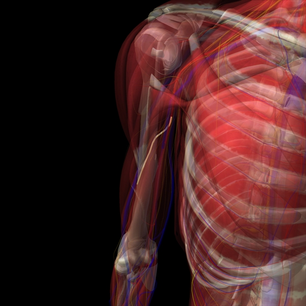
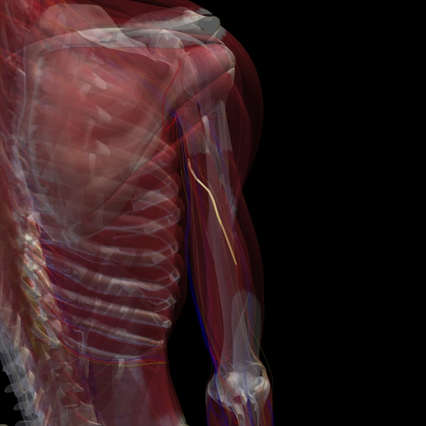
Note 1: Maybe better word instead rotation should be "pronation", but I am not sure. Pronation means turning back of forearm to front (supposed that hand is down beside body). I have seen a pronation movement on medical software, but this movement seemed to me unlikely, because only forearm turned, but the humerus bone of upper arm did not rotated. So in this article I suppose that humerus bone rotates when you try to give hand in takate kote position.
Note 2: This article concerns only one vulnerable area, that is placed just under Deltoid muscle. Another vulnerable area for TK positions is placed just above elbow, and you can identify it from medical/anatomy pictures.
For those who are interested on radial nerve location (concerning TK safety):
Scheme of Humerus bone - size cca 2 cm:
sizes of the humerus
View on non rotated arm, radial nerve:
"back" view on the arm
"back" view on the arm
"back" view on the arm 2
My schemes to illustrate how much the radial nerve "position changes" during 45° rotation:
Test of rotation of "arm" / humerus bone:
I took a pole and paper and draw the lines on it. According scheme that I have. The red line is radial nerve (a piece of wire that I turned around according scheme).
Rotated and non rotated:
Back side of shoulder would be on left and elbow on right side. These two images I use to paste them in schema, so that the lines that I have drawn, would fix schema.
Area located between black lines no. 2 and no. 3 would seem to me safe for face-down suspension, but because the arm have to be rotated 45° in Takate Kote position, then the radial position moves more up, too close to rope.
Non rotated:
Rotated:
Vertical:
Non rotated - rotated:
The dangers area no. 1
Results of the test:
This demonstration should show how much the radial nerve "changes position". The area from black line no. 2 to no 3. is the area I still speak about.
Result:
If you see the
vertical image - from the test is possible to see that the radial nerve appears to be close to right side.
The marked area black lines 2 - 3:
It seems to me that the
lower 1/2 is dangerous for face down suspension. In the very edge of the Deltoid it could be really dangerous to have ropes for suspension. Then,
in the upper 1/2 of marked area, it could be not so dangerous for F-D susp. but for safety reasons we should to place the ropes above this area.
For side suspension and back suspension the marked area in black lines 2 - 3 would be very dangerous area, where the ropes wouldn't be placed!
What danger?
The danger, that is related with incorrect rope placement or too tight tie, is the possibility that you can damage nerve. Too tight tie can cause nerve compression and this can hurt it. If we speak about radial nerve, which is placed in hand, then the injury causes impossibility to use toe or index finger. It can take few weeks to heal the nerve. To prevent this, we should place rope on body parts, which have muscles strong enough to prevent nerve damage. Rigger has to check ropes correct position and fixation or check it during the bondage play, if the tied person moves with ropes.