How to read diagram:
You need some image or arm with muscles like those medical images bellow. I looked for some way how to measure distances. I have found points which are approximately same distance from each other. These points are marked by long horizontal arrows filled with darker color. These are borders of the quarters. Use it as a rule, as if you would put 0 on the blue line. Then read it from bottom upwards. This is the way how I measured the distances. Notes in regular font are description if you go upwards. Notes in italic font are made for the case you read the diagram downwards, as in usual direction.
This diagram should be precise. It revealed some little mistakes in my previous estimations. I would dare to discuss this diagram with some medical educated person, but still be aware I am not a medic.

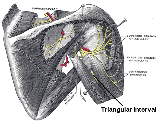
According other schemes of radial nerve, the path can vary. I have seen path going a bit more to right: in the triangular interval it turns right and does not enter tendon of the triceps. In that case, people with radial nerve like this, will be voluntary in the area between Deltoid and Triceps as I simplify to call it "valley". Also I have seen schemas, where tendon is shorter than this one and different schemas having either thinner or wider humerus.
Notes:
brachii - of arm
posterior - back
anterior - front
lateral - side
Note that at 2/4 and 3/4 radial nerve is covered by triceps medial head (great muscle). At the bottom of humerus it is covered by radiobrachialis muscle, which covers just little part of humerus on the left side of lateral view. You can use this diagram commonly with biodigitalhuman.com.
If you use Takate kote and you look for radial nerve path in takate kote, you must imagine that you rotate the humerus a bit towards right. I guess this rotation is about 20-30°, and can affect the look of the image just a bit.
PS:
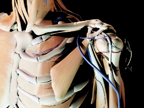
I add yet two images, there I tried to draw radial nerve according this diagram. I had to change proportion a bit because the humerus diagram was shorter, that model's upper arm.


You can also use this image to see the relation of position of Deltoid-Triceps-Lateral-Head valley and radial nerve located on humerus.
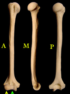
This source contains this image, showing cross-cuts of humerus, which is to show more exactly where the radial nerve (N. radialis) can lie:

You can see both views at same time: posterior and lateral. Anterior or back view is when you watch from bottom of the images upwards. Lateral or side view from outside to inside you get when you watch from right to left, and the image from profile.


